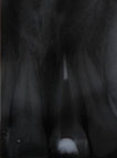
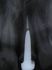
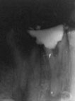
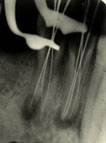
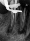
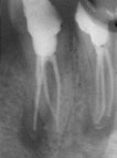
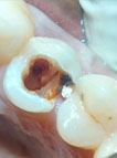
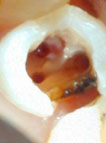
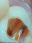
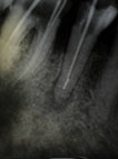
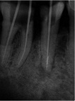
Custom Cast Post X ray shows tapering post space preparation. Apical 5mm GP retained for apical seal. Screw post in place. Lower premolar with 3 canals Diagnostic X – ray showing clinical variation in root morphology of lower pre-molar Three canals identified and working length measured Master cone image Post obturation image Perforation Repair Canals identified and enlarged. Perforation site cleaned with NaOcl and saline Perforation sealed and canal patency maintained Broken Instrument X ray showing broken instrument in apical third. GP stick stops at this point. Broken instrument is bypassed. GP positioned to full length of root.