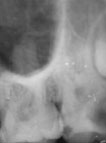
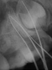
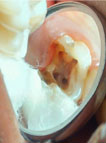
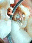
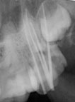
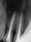
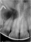
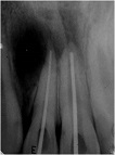
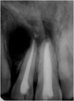
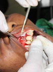
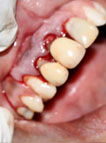
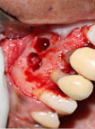
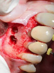
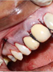
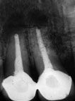
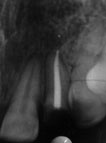
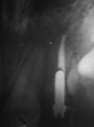
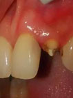
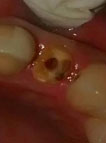
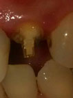
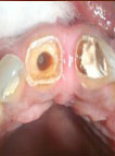
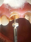
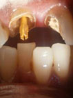
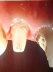
Upper 2nd molar with 4 canals Diagnostic IOPA Working Length Access view Master Cones in place Master cone X-ray Post Obturation View Non surgical Management of Leison Case 1 Pulpa irritants are removed from canals and working length determined Wein’s recommendations are followed and master cone fit checked When the canals are dry and ready for obturation they are sealed as per lateral condensation technique Case 2 Pre Operative Master Cones in place Post Obturation View Surgical management of Lesion Working length file is used to measure length of vertical releasing incision Crevicular incisions and single vertical releasing incision planned. The lesions are exposed and curetted The files exit through root tip The root tips are prepared, the flap is repositioned and sutures are placed Post op radiograph Tapered Metal Screw Post (Case I, Case II, Case III) Case I Radiograph checked for apical seal, thickness of dentin, and root length. Post placed to 2/3rd of root length. Broken tooth. Post space prepared. Screw post in place. Case II Post space shows condensed Gutta Percha. Peeso reamer inserted to verify post space preparation and length. Post placed and checked for occlusal clearance. After coronal built up.